Search This Blog

Tuesday, March 30, 2010

Organ transplantation

TRANSPLANTATION TRANSPLANTATION 
The replacement of tissue or organ either in same or in different organism is called transplantation. This process is used to replace damaged or diseased tissue or organ of the body. 

TYPES OF TRANSPLANTATION: 
On the basis of the relationship of donor and recipient, transplantation is classified into the following type: a)AUTOGRAFT: 
The transplantation in which an isolated portion of living tissue is taken from one part of body and transferred to another part of same individual is called Autograft. Eg: skin graft used for severe burnt skin 

b)ISOGRAFT: 
The transplantation between genetically identical individual is called isograft. Eg the grafting between twins. This type of transplantation is the most successful as donor and recipient are genetically identical. 

c)ALLOGRAFT: 
The transplantation between two individual of same species but with different genetic background. Eg:a heart or kidney transplantation 

d)XENOGRAFT: 
The transplantation between individual of different species. Eg: monkey to man 

ORGAN TRANSPLANTION: 
The replacement of complete organ like kidney, heart ,lung etc. between two individual is called organ transplantation. It is a decisive procedure used in those cases where survival of the animal is in danger. For success donor and recipient must be genetically identical. Organ transplantation is great achievement in medical science. Kidneys have been transplanted for several years with varying degree of success. The first heart transplantation was done in 1967 by Christian Barnard in the USA and since then attempts have been made to graft lungs and liver. The main problem in organ transplantation is that the recipient body recognizes transplanted organ as foreign body or antigen. Therefore, the recipient body produces antibodies against grafted organ. Thus, a foreign organ graft will be rejected by the recipients immune system. This is called rejection. The best known example of organ transplantation is the transport of cornea because cornea does not link with the immune system of the body as well as with the blood supply. Its transplantation is also relatively simple. However, transplantation of other organs like kidney, liver, heart etc. is very complicated as it is not accepted by the human immune system. 

WAY TO PREVENT REJECTION:
1)By using immuno suppressive drugs. These drugs are immuno suppressants. These drugs are very expensive and make the body highly susceptible to infection.
2)Transplantation should be between genetically identical individual like twins. 
3)Immune system can be suppressed by radiation. 
4)Research is still on to develop immuno suppressive drugs which only stop rejection response while the rest of the immune system may remain active to fight other invaders. 
5)Now a days, research is going on for the development of artificial organ as liver and heart. Transplantation of artificial organ is most encouraging. 

ADVANTAGES OF ORGAN TRANSPLANTATION: 
1)Organ transplantation helps in the replacement of damaged or diseased tissue or organ from the body. 2)Organ transplantation is decisive procedure used in those cases where survival of animal is in danger. 3)Organ transplantation is great achievement in medical science. 
4)Organ transplantation is boon to save the life of patients from organ failure. 
5)Organ transplantation is possible to grow sheet of skin from a small piece of skin.This has led to establishment of tissue bank. 
6)Cornea transplantation is are done to restore vision. Bone marrow is transplanted to treat leukemia and disease of blood cells.

Kreb cycle and Glycolysis

TYPES OF RESPIRATION: The oxidation of organic substance in living cells resulting in the liberation of energy is called respiration. It is of two types: 
1)AEROBIC RESPIRATION: 
This is known to occur in presence of oxygen and is described as a vast series of enzymatic reaction through which the chemical energy contained in the food is converted into energy in the form of ATP. The energy contained in food is made available for work through glycolysis and Krebs's cycle. The glucose is oxidized to its component CO2 and H20 with the release of energy in the form of ATP. The overall reaction is represented as: 
C6H12O6+ 6O2--------6CO2 +6 H20 +Energy 
The first phase of glycolysis is known to occur in cytosol of the cell where the Krebs cycle is known to take place in mitochondrial matrix. 

GLYCOLYSIS: 
Glycolysis is described as series of enzymatic reaction using glucose as raw material. It is also alternately termed as Embden-Mayerhof-Parnas(EMP) after the name of three German Plant Physiologist who work on this path way. 
1)Initially the glucose molecule accept the phosphate group of ATP and is converted into Glucose 6 phosphate in presence of Enzyme Hexokinase. 
2)The glucose 6 Phosphate accept the phosphate group of ATP and is converted into Fructose1,6 diphostate which is 6 carbon containing compound in presence of enzyme Phospho fructose kinase. 
3)The fructose 1,6 diphosphate is cleaved by the enzyme aldose into 3 phosphoglyceraldehyde(PGAL) and Dihydrooxyacetonephosphate(DHA
ATP'S IN KREB CYCLE AND GLYCOLYSIS
P).  
The DHAP is converted into its isomer 3 PGAL by enzyme phosphotriose isomerose.The 3-PGAL accepts on phosphate group from inorganic phosphate and is converted into 1,3 diphosphoglyceric acid(1,3di PGA) which donates a phosphate group to ADP and is 3Phosphoglyceric acid.In this process,a molecule of ATP is generated and the enzyme involved is PHosphoglyceric acid kinase. Intramolecular transfer of the phosphate group occurs from the 3 rd carbon of PGA to 2nd carbon therby forming 2-PGA in presence of enzyme Phosphoglycero mutase. 2-PGA loses one molecule of water and is converted into Phosphoenol Pyruvic acid(PEPA) with the involvement of enzyme enolases. The PEPA donates one phosphate group to ADP and is converted into pyruvic acid.In this process one molecule of ATP is generated and the enzyme involved is pyruvate kinase. ENERGETIES OF GLYCOLYSIS: 1)No of ATP's consumed-2 ATP 2)No of ATP's generated a)1,3 diPGA----3PGA ----1 ATP b)PEPA-----Pyruvic acid----1 ATP 1X2=2 c)Oxidation of NADH2=3 ATP 3X2=6 ATP Total ATP's generated-----10 ATP KREBS CYCLE: The 2nd phase of the oxidation of food involves the Kreb's cycle or Tricarboxylic cycle(TCA).It is series of chemical reaction by which the pyruvic acid produced is completely broken down into CO2 and water with the uptake of O2 and release energy in the form of ATP. The complete pathway was worked out by Sir Hans E Kreb(1953) for which he was awarded the nobel prize in the year 1953.All the reaction of this cycle occur in the mitochondrial matrix. For the participation of pyruvic acid in the Kreb cycle ,it is initially converted into Acetyl coenzyme A by the process of oxidative decarboxylation which can be summed up as Pyruvic acid + coenzymeA +NAD------Acetyl CoA +NADH2 + CO2 The acetyl CoA reacts with Oxalo ACetic Acid and water to form Citric acid in presence of enzyme citric acid asynthese. Acetyl CoA +OAA+ H2O------Citric acid+ co enzyme The citic acid loses one molecule of water to form sis aconotic acid which react with water to form isocitric acid. CA + excess H2O-----Sis Aconitic acid+H2O-----Isocitric acid The iso citric acid is dehydrogenated by the enzyme to form oxalo succinic acid. `ICA +NAD---------Oxalo Succinic acid+ NADH2 dehydrogenase (OSA) The OSA loses one molecule of CO2 to form alpha ketoglutaric acid. OSA-----------------------alpha ketoglutoric acid + CO2 decarboxylase The alpha ketoglutaric acid undergoes oxydate decarboxylation to be converted into Succinyl CoA. alpha ketoglutaric acid+CoA +NAD-------------------------Succinyl CoA alpha ketoglutoric acid dehydrogenase The succinyl CoA loses the CoA to be converted into Succinic acid. Succinyl COA +H2O +H3PO4-------Succinic acid +Co A+GTP(Guanoshine triphosphate) sucinyl thiokinase (GTP+ADP----ATP) The succinic acid is dehydrogenated into fumaric acid which react with one molecule of water to from malic acid. SA+FAD-------------------fumaric acid +H2O-----------malic acid(MA) Dehydrogenase The MA is dehydrogenated to form OAA by the enzyme malic acid dehydrogenase. MA+NAD--------------OAA+NADH2 ENERGETICS OF KREB CYCLE: 1)In the conversion of PA to Acetyl CoA 3ATPs( 3X2=6) 2)Iso CA------OSA 3ATPs( 3X2=6) 3)alpha keto glutaric acid------succinyl COA 3ATPs( 3X2=6) 4)SA------FA 2ATPS(2X2=4) 5)MA----OAA 3ATPs( 3X2=6) 6)Succiny COa------SA 1ATP(1X2=2) TOTAL ATP'S IN KREB CYCLE=30 ATPS TOTAL ATP'S PRODUCED FROM ONE MOLECULE OF GLUCOSE=40 ATPS TOTAL NO OF ATPS CONSUMED=3 ATPS NET GAIN OF ATPS FROM ONE MOLECULE GLUCOSE=38 ATPS

Respiration in human beings

RESPIRATION:VOLUME OF AIR
RESPIRATION IN HUMAN BEINGS:
Respiration is process of taking in oxygen to oxidise the food and release carbondioxide,energy and water.Energy is used to perform metabolic activities whereas carbondioxide and water is exhaled during expiration.
RESPIRATORY TRACT:
Respiratory tract includes the passage through which air reach up to lungs.Respiratory tract consists of following part:
1)External nares:
The external nares are a pair of slit at the lower end of the nose.They open into the nasal chamber.Air enters into the nasal chamber through these nares.
2)Nasal chamber:
The nasal chambers are the pair of passage in the head above the palate.Each chamber show three region.They are:
a)Vestibular region:
It has hair which filter the air.
b)Respiratory region:
it is thin walled and highly vascular.It makes the air warm.It also bear mucus gland which secrete mucus.
c)Olfactory region:
It detect the smell.It open into internal nare.
3)Internal nares:
The nasal chamber open into nasopharynx by internal nares.The inhaled air passes into nasopharynx through internal nares.
4)Pharynx:
The pharynx is short funnel like tube behind the buccal cavity.The food and air passage cross here.Its upper part is nasopharynx in which the internal nares and eustachian tube open.The middle part is oropharynx which receives the buccal cavity.The lower part is laryngopharynx.It leads to two tubes :the one in front is wind pipe or tarchea and the one at the back is the food pipe or osopharynx.
5)Larynx:
larynx is the upper part of the trachea.It is short,tubular chamber which is often called Adam's apple or voice box.
6)Trachea:
Trachea is a long tube which is 25 cm long and 2.5cm in diameter.It runs through the neck and enters into the anterior region of thorax and divide into right and left bronchus.Trachea and bronchus are supported by incomplete cartiliginous ring.It provide free passage to air.
7)Bronchus:
Each bronchus open into lung.After entering into lung,it divide into primary,secondary,tertiary and finally into bronchioles.Bronchioles open into terminal duct which open into alveoli.Each alveolus is surrounded by cappilaries of vein and cappilaries of artery for exchange of gases.Bronchial tree include the bronchus which divide repeatedly inside the lung and end into alveoli.
8)Repiratory organ:The lung
Lung is spongy,pink colour moist organ which lie in anterior thoracic cavity inside the pleural cavity.It is covered by double layered pleural membrane.Between the pleural membrane is pleural fluid which keep the lungs moist and reduces friction.The left lung is slightly smaller than left lung.Between the left lung and right lung there is a space known as mediasternum.Towards the posterior end of left lung has cardiac notch which provide space for heart.The left lung is divided by oblique fissure into anterior lobe and small posterior lobe.In right lobe the horizontal and oblique fissure divide into anterior lobe,posterior lobe and median lobe.The lung is completely filled by alveoli.Each alveolus is surrounded by cappilaries of vein and cappilaries of arteries.
MECHANISM OF LUNGS:
Mechanism of lungs include mainly two process and they are inspiration and expiration.Inspiration is taking in of oxygen to lungs and expiration is releasing carbon dioxide from lungs to external environment.
The lung is located in thoracic chamber which has dorsally verterbrae,ventrally sternum and on the sides has rib.Anteriorly vertebrae and sternum is joinged;and posteriorly there is dome shaped diaphragm.Between the rib has two set of muscle i.e external intercostal muscle and internal intercostal muscle.Conraction and relaxation of intercostal muscle increase and decrease thoracic valve.
1)Inspiration:
#During inspiration,external intercostal muscle contract and internal intercostal muscle relax so that ribs move forward and downward.
#Due to movement of ribs,sternum move downward so that dome shaped diaphragm stretch and become flat.
#So,that the thoracic volume is increased and lungs expand.
#The air pressure inside the lung is lower than external environment.
#Due to difference in air pressure ,the air rush from external environment into lung through respiratory tract where gaseous exchange take place.
2)Expiration:
#During expiration,internal intercostal muscle contract and external intercostal muscle relax so that the ribs move backward and upward.
#Due to movement of ribs,the sternum move upward and return to original position.
#Due to this the diaphragm become dome shape.
#Thoracic volume decreases and increases pressure in the lungs.
#As a result,air pressure inside the lung is higher than that of external environment.So,the air rush outside from lung to external environment.
PULMONARY AIR VOLUMES:
The amount of air exchanged during breathing is measured by appratus called Spirometer.The quantities of air the lung can receive,hold or expel under different conditions are called pulmonary volumes.The following are the terms used to express pulmonary volume:
1)Tidal Volume(TV):
It is the volume of air that is breathed in and out during effortless normal breathing.In a normal adult person this volume is about 500ml.
2)Inspiratory Reserve Volume(IRV):
It is the total volume of forced inspired air inn addition to normal inspired air(tidal volume).It is approximately 1500-2500ml.
3)Expiratory Reserve Volume(ERV):
It is the total volume of air expired forcefully in addition to normal expired air.It is approx. 1500ml.
4)Vital Capacity:
It is the volume of air,which can be maximum inspired and also maximum expired.It is about 4500ml in males and 3000 ml in females.
Vital capacity=IRV+ERV+TV
5)Residual Volume:
The volume of air that remains in the lungs after maximum forced expiration is called residual volume.It is about 1500ml.
6)Total lung capacity:
The volume of air in lungs and respiratory passage afer a maximum inspiratory effort is called total lung capacity.
Total lung capacity=vital capacity + Residual volume
7)Dead space:
It is the volume of air trapped in respiratory passage during inspiration is called dead space.It is about 150 ml
8)Respiratory Quotient:
It is the ratio of carbon dioxide evolved to oxygen absorbed.Some of the value of respiratory quotient of subsatances are:
carbohydrate:1
Protein :0.9
fat :0.7

PHYSIOLOGY OF RESPIRATION:
1)EXTERNAL RESPIRATION:
The exchange of Carbondioxide and oxygen between fresh air of alveolus and carbondioxide containing blood of alveolar cappillary by diffusion is called external respiration.It takes place mainly due to partial pressure of Carbon dioxide and oxygen.

OXYGEN TRANSPORT:
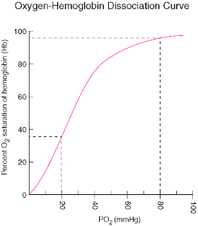
3% of oxygen is carried by blood plasma whereas 97% of oxygen is carried by Haemoglobin as Oxyhaemoglobin.When the partial pressure of oxygen is high,more oxy hRESPIRATION:VOLUME OF AIRaemoglobin is formed.15 gm of Hbis present in 100ml of blood.One molecule of iron of haemoglobin can bind four molecules of oxygen.the amount of oxy haemoglobin formed is called percent saturation of Haemoglobin.
The relation between percent saturation of haemoglobin and Partial pressure of oxygen in mm is expressed by dissociation which is of sigmoid shape.
When the partial pressure of oxygen is 70 mm Hg the blood bceome completely saturated with the oxygen.When the partial pressure of oxygen decreses that means partial pressure of carbondioxide increases the oxyhaemoglobin dissociates into haemoglobin and oxygen.Therefore the partial pressure of carbondioxide is directly pressure to percent dissociation of oxy haemoglobin.This is known as Bohr's effect.

2)INTERNAL RESPIRATION:
By the dissociation of oxy haemoglobin,free oxygen is produced which diffuses into cell, tissue or organ due to concentration gradient.The oxygen is used to oxidise the food to give energy,carbon dioxide and water.This is known as internal respiration.Energy is used to perform different metabolic activity of body whereas carbon dioxide and water is carried by blood in different form upto alveolar cappilaries.

CARBON DIOXIDE TRANSPORT:
Blood carry carbon dioxide and water in different forms upto alveolar cappilaries:
1)CO2 TRANSPORT AS CARBONIC ACID(H2CO3):
5-10% Of carbon dioxide is carried by bllod plasma as carbonic acid .
H20 +cO2----H2CO3
The respiratory enzyme carbonic anhydrase acts as catalyst during the formation of carbonic acid.

2)CO2 TRANSPORT AS CARBAMINO COMPOUND:
About 15% of CO2 is transported as carbamine compound.Amino group of haemoglobin also carbon dioxide in the form of carbamine compound upto alveolar coppilaries.
3)ABOUT 30-35% OF co2 IS CARRIED AS NaHCO3 and KHCO3:
RBC carry carbondioxide in the form of potassium bicarbonate(KHCO3) and plasma carry carbon dioxide in the form of sodium bicarbonate(NaHCO3).There is the excess carbon dioxide and water which diffuse in RBC and makes carbonic acid(H2CO3).As the RBC can not carry carbonic acid,so it dissociates into H+ and HCO3-.
Excess CO2 + H2O---------H2CO3-------H+ + HCO3-
diffuses
Now the potassium haemoglobin(KHb)begins to dissociates into K+ ion and Hb.K+ ion together with HCO3- ion makes KHCO3 and H+ion together with haemoglobin makes KHb.Excess KHCO3 begins to dissociate K+ ion and KCO3- ion.The HCO3- diffuse from RBC into plasma because RBC is permeable to negatively charged ion.Plasma has NaCl.Due to the deposition of HCO3- ion,NaCl dissociate into Na+ and Cl-.The Na+ together with HCO3 make NaHCO3.To make ionic balance,chloride shift from plasma to RBC and with K+ makes KCl.
The mutual exchange of Cl- and HCO3- between plasma and RBC to make ionic balance is called chloride shift.In this way,NaHCO3 and the KHCO3 is carried by plasma and RBC to alveolar cappilaries.The HHb dissociates into H+ and Hb.The KCl also dissociates into K+ and Cl-.Now the K+ with Hb makes KHb so that Cl- shift back into the plasma.Now,NaHCO3 dissociates into Na+ and HCO3-.SO,Na+ and HCO3-.SO,Na+ with Cl- makes Nacl.Hco3- also move back to RBC and with H+ ion make H2Co3.Similarly,H2CO3 dissociate into water and carbon dioxide which diffuse from alveolar cappilaries into alveoli and exhaled during expiration.

MIXTURE:SYNAPSE AND NEUROTRANSMITTER:

MIXTURE:SYNAPSE AND NEUROTRANSMITTER:
SYNAPSE AND NEUROTRANSMITTER:
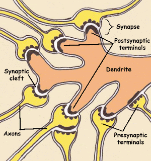
There is always involvement of more than one neuron for the transmission of nerve impulse.It might be the sensory or motor.At the free end of axon,the axon branches as well as the terminal end swells.These swellings are known as Synaptic knobs or buttons.There is no physical contact between the two nerve cell that involve in action potential.The gap that is formed between pre synaptic neuron and post synaptic neuron is called Synaptic cleft and the process is called Synapse.
The synapse is an area of functional contact between one neuron and another for purpose of transferring information.Synapses are usually found between the line terminal branches of axon of a neuron and the dendrites or cell body of another.This type of neuron is called Axon dendrite synapse.Sir Charles Sherrington was the first person who used the term 'synapse the junction point between two neurons.
In synaptic knob,Vesicles that contain neurotransmitter are present.Specific neurotransmitter are released by exocytosis from presynaptic neuron which diffuses with dendrites or cell body of post synaptic neuron.These chemicals help in the transmission of nerve impulse.The dopamine,histamine,serotonin,adrenalin,nor adrenalin,gamma amino butalic acid(GABA) are the neurotransmitter of nervous system.

NERVE IMPULSE CONDUCTION:

Nerve impulse is defined as the sum total of physical and chemical disturbances created by stimulus in the nerve cell.It is also defined as electrochemical event that changes membrane potential of nerve fibre and causes transmission of stimulus from receptor to CNS and vice versa.
An impulse is initiated by stimulation of sensory nerve ending or by the passage of an impulse from another nerve.Transmission of impulse or action potential is due to movement of ions across the nerve cell membrane.During rest,the nerve cell membrane or axolemma is polarised due to the difference of concentration of ion across the membrane.This means that their is different electric charges on either side of membrane.This is called resting membrane potential.The charged electric potential of axolemma is called action potential.The main ions involved in nerve impulse conduction is Na+(main extracellular cation)and k+(the main intracellular ion)
In resting state the sodium are actively transported from axoplasm to extracellular fluid as a result the concentration of sodium inside or in axoplasm is almost negligible.During this period the concentration of sodium is 100 times more than that of axoplasm.This is due to the active movement of sodium and is termed as Na pump.When the stimulated Na pump stops and the permeability of axolemma inceases by which Na+ diffuses from extracellular fluid to axoplasm.This cause depolarisation.Dipolarisation is very rapid enabling the conduction of nerve impulse along the entire lenght of neuron in a few millisecond.
In myelinated neuron,the insulating properties of myelin sheath prevent the movement of ion.THerefore electrical charges across the membrane can only occur at node of Ranvior.When an impulse occur at one node ,dipolarisation passes along next node so that the flow of current appear to leap from one node to another.This is called Saltatory conduction.Myelinated fibre conduct impulse faster than unmyelinated fibre because saltatory conduction is faster than complete conduction.The fastest fibres can conduct impulses at the rate of 130m/s as in skeletal muscle while the slowest impulses travel at 0.5m/s,

Monday, March 29, 2010

Plant Breeding and Hybridisation

PLANT BREEDING

PLANT BREEDING: 

 Plant breeding is a process of developing improved varieties of plants with desirable characters from the existing ones is called plant breeding. It can also be defined as the improvement of plants by the application of genetic principles. 

METHODS USED IN PLANT BREEDING: 1)PLANT INTRODUCTION: 
 It is a process of introducing new plants with desirable characters from their area of origin to a new area is called plant introduction. This process is very fast and easy method of breeding. If the plant if brought from foreign country than it is called exotic plant and if it is from the same country then it is called as indigenous plant. Adjustment of plants to new or changed environment is called acclimatization. Plant introduction are usually done by cuttings or seeds. 

2)SELECTION: 
 It is the oldest method of breeding process. It involves picking up of better ones from the entire crop plants. 
TYPES OF SELECTION:
 a)MASS SELECTION: 
 It is the simplest method followed by the farmers to improve mainly local varieties of crops. Before harvesting the crops, best looking and healthy plants are selected from the mass. Seeds of these plants are collected and sowed them to raise new generation. Mass selection is based on external character. b)PURELINE SELECTION: 
 A pure line is progeny obtained from a single individual by self fertilizations. A group of plants obtained from a single self fertilized homologous plant is called pureline. It involves a desirable homologous individual from the mixed population and multiplying the same. Pureline selection is the only method of improvement of local varieties of self pollinated crops. The progeny of pureline selection are similar in phenotypical and genotypical character. 
c)CLONAL SELECTION: 
 A clone is defined as a progeny of a single plant obtained by vegetative propagation. The clonal selection is concerned with the selection and propagation of best individual of clones from mixed population of vegetative propagated plant. 
 
3)HYBRIDISATION: 
 The process of crossing two or more plants together to get offspring of new desirable characters as a result of genetic recombination is called Hybridization. 
TYPES OF HYBRIDISATION: 
a)INTRASPECIFIC HYBRIDISATION:
Crosses are made between two individuals of same species. This crosses are done to improve self pollinated crops producing homozygosity or pure offspring. 
b)INTERSPECIFIC HYBRIDISATION: 
 Crosses are made between individual of different varieties of the same species. This crosses are made to improve self pollinated and certain cross pollinated crops.
c)INTRAGENERIC HYBRIDISATION
 Crosses are made between two individuals of different species belonging to same genera.it is used to produce resistance varieties from diseases ,frost or drought.
d)INTERGENERIC HYBRIDISATION
 Crosses are made between two individuals of different genera belonging to same family. It produces resistance varieties as well as desirable combination of all characters. Eg:Brassica X Raphanus=Raphanusbrassica 
 
TECHNIQUES FOR HYBRIDISATION: 
 It is necessary for the breeder to have following information: 
a)Whether plants are self or cross pollinated. 
b)Whether plants are monoecious or dioecious.
c)Whether the flowers are unisexual or Bisexual. 
d)time of Anthesis. 

 PROCESS OF HYBRIDISATION: 
a)SELECTION OF PARENTS: 
 Male and female parents, with desirable characters are selected from the available materials. Both the parents should mature at the same time. 
b)EMASCULATION: 
 Removal of anthers from bisexual flowers before they dehisces or shed their pollen is known as emasculation. It is done in order to prevent self fertilizations. Emasculation is not necessary in the parents are monoecious. 
c)BAGGING:
After emasculation, flower buds are kept enclose in bags made up of cloths, plastic or polythene etc. It is done to prevent pollination through unknown pollen. 
d)CROSSING: 
It is an artificial pollination. Pollens are collected from the desirable male flowers. These collected pollens are then dusted on the stigmas of pistils. After crossing the female flowers are bagged again. 
e)LABELLING: 
The female parents are then labelled properly. The labelling should bear the following information. i)Serial number ii)Details of male parents and female parents. iii)Date of emasculation and crossing 

f)HARVESTING: 
 At the end of the season, the fruits and seeds of each cross are separately collected. They are dried and preserved and then sowed to raise the F1 generation. 

HYBRID VIGOR: 
 The superiority of offspring's over their parents is known as hybrid vigor or heterosis. 

 4)MUTATION BREEDING; 
 Mutation is define as a sudden change in hereditary of an organism. They may be due to changes in chromosome number(Polyploidy),changes in the structure of chromosome(chromosomal abbreviation) or changes in molecular level of DNA(gene mutation). Mutation may occur naturally or artificially, produced by exposing organism to mutagenic agents like X-rays, Gamma rays etc. 

5)POLYPLOIDY BREEDING: 
 Polyploidy breeding involves use of polyploidy in crops improvement. In general the odd numbered polyploidy 3x,5x etc. are sterile while even numbered polyploid are fertile. There are two main categories of polyploidy. 
a)Autopolyploid(multiples of same genomes) b)Allopolyploids(multiples of different genome) 

 APPLICATION OF PLANT BREEDING: 
 The main objective of plant breeding is to produce improved variety of plants with desirable character from existing ones based on principles of genetics. Some of the application of plant breeding are listed below: 
a)INCREASE IN YIELD: 
Nowadays, plants breeders have become successful in producing improved varieties of crops that can produce higher yields of cereals, fibers, oil, sugar etc. 
b)IMROVED QUALITY: 
 Through plant breeding, good quality of plant with desirable character are produced. 
c)RESISTANCE OF DISEASE: 
 Every year thousands of plants get affected by several diseases. But plant breeding has enabled to produce disease resistance varieties plants. 
d)PRODUCTION OF VARIETIES SUITED TO PARTICULAR CLIMATE AND SOILS: 
 Plant breeders are able to develop hardy varieties which can withstand drought and frost. 
e)CHANGE IN DURATION OF MATURITY: 
 Plant breeding has shortened the duration of crop maturation as result of which more types of crops are produced in same year. 
f)RESISTANCE TO LODGING: 
 Plants breeder are producing plants with between lodging.

Plant tissue culture

Plant tissue culture
PLANT TISSUE CULTURE:
The process by which desirable plants can be grown from any plants part,tissue or cells artificially in the laboratory in an artificially prepared nutrient medium under aseptic condition is known as plant tissue culture.

HISTORICAL BACKGROUND OF TISSUE CULTURE:
Haberlandt(1896) was the first person to culture isolated vegetative cells.He was able to maintain the cell in the medium but failed to differentiate it.In 1934 different worker P.R. white,R.s Gautherate and P.Hobegurt were able to grow cambium cells from tobacco stem and carrot root on artificial culture medium.However,this culture failed to differentiate and grew as undifferentiated masses of parenchymatous cells called callus which could be propagated indifinitely by repeated subculturing on fresh culture medium.

EQUIPMENTS FOR SUCCESS OF TISSUE CULTURE:
To get success in vitro culture or micropropagation a laboratory should have following equipments and facilites.They are:
i)pH meter ii)Chemical balance iii)Hot air oven iv)Centrifuge v)Auto clave vi)UV lamp vii)Shaker
viii)Dissecting microscope ix)Compound microscope x)Refrigerator xi)Laminar air flow cabinet xii)Essential glass

METHOD OF TISSUE CULTURE:
a)NUTRIENT MEDIUM:
Cultured tissue can not synthesize their own food and need an external supply i.e they are heterotroph.The medium or cultured medium used in tissue culture is basal medium.It include following things:
i)INORGANIC NUTRIENTS:
It includes all the 16 elements which are essential for normal and healthy life of plants.These elements can be grouped into Macro and micro elements.
MACRO ELEMENTS: C,H,O,P,K,Ca,S,Mg
MICRO ELEMENTS:Zn,Fe,Cu,B,Mn,Mo and Cl
ii)ORGANIC ELEMENTS:
It includes sucrose,glucose,fructose,carbohydrats and vitamins.
iii)NATURAL EXTRACT:
natural extract like yeast extract,coconut milk,tomato juice,malt extract are added in the medium.
iv)GROWTH HORMONE:
it includes auxin,cytokinin and gibberellin
Some of the standard media available are Murasbige and Skoog's media,White's media and Nitschs media.
v)AGAR:
Agar is a polysaccharide substance obtained from sea weeds which is used to provide solid surface for growth.

b)STERILIZATION OR ASEPTIC CONDITION:
The culture must be totally free from microbian contamination.Microbes may enter culture through the ingredients of medium,througn plant organ or explant and through air so that the culture vessel and instrumenti.e glasswares,metal instruments are sterilized by exposure to hot dry air at 160 C to 170 C for 2-4 hours in a hot air oven.
i)CULTURE MEDIUM:
Culture vessels containing the medium are plugged and autoclaved at 120 C for about 15-20 minutes.
ii)PLANT MATERIAL OR EXPLANT:
Plant material or explants are surfacesterilized by using sodium hypochloride or calcium hypochloride solution.After surfacesterilization plant materials are washed 3-4 times in a sterile distill water.
iii)TRANSFER AREA:
Inoculation is carried out in a laminar air flow cabinet.In this cabinet filterate sterile air flows inside at a constant rate.The flow is unidirectional and makes the cabinet sterile.
To avoid contamination hands and arms are washed with soap and then 95% ethanol.Thus an aseptic environment is maintained for tissue inoculation.

c)LIGHT:
Normally it is not necessary for growth of culture but it plays an important role in inducing differentiation.The intensity and duration of illumination varies from species to species.

d)TEMPERATURE:
Generally 25 to 27 degree centigrade is necessary for callus growth.

e)HUMIDITY:
A relative humidity of 70-75% is optimum for the growth of culture.

TYPES OF PLANT TISSUE CULTURE:
Plants material used for plant tissue culture is known as explants.On the basis of explants used for plant tissue culture,it is of following type:
a)SHOOT CULTURE:
Plant tissue culture in which sterile shoot tips or axillary buds are used as explants is called Shoot culture.

b)PROTOPLAST CULTURE:
Here,protoplast is used as culture.Somatic hybrid can be produced from protoplast culture.

c)EMBROYO CULTURE:
The plant tissue culture in which embroyo is used as explant is known as embryo culture.

d)ANTHER CULTURE:
The plant tissue culture in which anther is used as explant is known as Anther culture.By this haploid plant is produced which is of great importance to scientist as mutation can be induced in them.

e)MERISTEM CULTURE:
The plant tissue culture in which apical meristem is taken as explant is known as meristem culture.Through this disease resistance plant can be produced.

APPLICATION OF PLANT TISSUE CULTURE:
1)MICRO PROPAGATION:
Rapid asexual or vegetative propagation of plant in vitro is called micro propagation.Large no of plants can be produced throughout the year.

2)SOMATIC HYBRIDIZATION:
Fusion of somatic cells in vitro is called somatic hybridisation.Novel hybrid can be produced in sexually incompatible species.

3)PRODUCTION OF HAPLOID PLANTS:
Through anther culture haploid plants are produced.It is very important in research point of view as mutation can be induced and detected.

4)PRODUCTION OF PATHOGEN FREE PLANT:
Through meristem culture,virus free plants can be produced from diseasesd material.

5)PRODUCTION OF DISEASE RESISTANT VARIETIES:
Many plants are dying due to presence of virus or bacteria.So,plant tissue culture has been able to produce disease resistance variety of plants.

6)MINIMIZE THE USING SPACE:
Tissue culture can be used to minimize the growing space in commercial nurseries for maintainance of stock plants.

TERMS:
CALLUS:
Callus is mass of vacuolated,unorganized and undifferentiated cells prepared in artificially cultured medium in aseptic condition in lab from a cell or tissue in plants.

Glands:Pituitary and thyroid gland

Glands are the groups of specialized cells that produce some chemical substance needed in Metabolic processes of the body. 

TYPES OF GLANDS: 
1)EXOCRINE GLAND: 
The glands whose secretion are carried by ducts into the site of action is known as exocrine gland.They are present near the site of action of their secretions. They produce enzyme. Eg:Pancreas,Salivary gland 2)ENDOCRINE GLAND: The ductless glands whose secretion are directly poured into the blood is known as endocrine gland.They are present at sites distant from sites of action of their secretion.They produce hormones.Eg:Pituitary gland,Thyroid gland etc. The main function of endocrine system is to maintain homeostasis withe the help of autonomic nervous system. 

ENDOCRINOLOGY: 
The study of endocrine glands and its secretion is known as endocrinology. THOMAS EDDISON is regarded as Father of Endocrinology. HORMONES: Hormones are the chemical messengers secreted by endocrine glands that are circulated in blood and act on specific target cells to bring about the biological effect. 
TYPES OF HORMONES: 
1)AUTOCRINE HORMONE: 
Hormones that act on the cells secreting it is called autocrine hormone. Eg:Platelet activating factor 2)PARACRINE HORMONE: Hormones that act on the cell adjacent to the cell secreting it is called paracrine hormone; also called local hormones. Eg:gastrin,secreted by G cells act on the parietal cell of the stomach to secrete HCl. 

CHEMICAL CLASSIFICATION OF HORMONE: 
1)AMINE HORMONES: Derived from amino acid tryosine.Eg:Thyroid hormone, epinephrine, norephinephrine,dopamine 
2)STEROID HORMONE: Derived from cholesterol.Eg:Sex hormone and adrenal cortex hormones 3)PEPTIDE HORMONE: Eg:pancreatic hormone,pituitary hormone,parathyroid hormone,calcitonin

TYPES OF ENDOCRINE HORMONE: 
1)PITUITARY GLAND/HYPOPHYSIS: Pituitary gland is a small pea shaped,reddish grey endocrine gland that lies at the base of the brain below hypothalamus.It is also known as master gland as it controls activities of almost all endocrine gland and effect metabolism of somatic gland.Pituitary gland is connected to the base of hypothalamus by a small stalk called pituitary infundibulum.Pituitary gland along with hypothalamus is known as hypophysis as it lie in hypophoseal fossa of sphenoid bone.It consists of three parts: 
a)ANTERIOR LOBE: It is important part of Pituitary gland which secrete eight different hormone that stimulate the glands.
Glands:Pituitary and thyroid gland HORMONES SECRETED BY ANTERIOR LOBE: 
i)FOLLICLE STIMULATING HORMONE(FSH): FSH stimulate the development and maturation of ovarian follicles which produce egg in female and promote spermatogenisis in male. 
ii)LEUTINIZING HORMONE(LH): LH stimulate ovulation,formation of corpus luteum and production of secondary growth hormonei.e progesterone and oestrogene. In male,LH is known as interstitial cell stimulating hormone(ICSH).It stimulate interstitial cell of testis and produce testerone.
iii)PROLACTIN HORMONE(PH): It stimulates lactation(milk production) in female. iv)ADRENOCORTICOTROPHIN HORMONE(ACTH): It stimulate adrenal cortex and produce adrenocorticosteroid hormone. 
v)THYROOXINE OR THYROID STIMULATING HORMONE(TSH): It stimulate thyroid gland. vi)GROWTH STIMULATING HORMONE OR SOMATROPHINE: It stimulate growth hormone.Deficiency of it causes Dwarfism in child and hypersecretion leads to gigantism in child and Acromegaly in adult. 

b)INTERMEDIATE LOBE: It arises from connective tissue.
 i)MELANOCYTE STIMULATING HORMONE: It stimulate pigmentation of skin. 

C)POSTERIOR LOBE: It is down growth of nervous tissue of hypothalamus.It collects the hormone secreted by neurosecretory cells of hypothalamus. i)OXYTOCIN: It causes contraction of smooth muscle during child birth.It also stimulate milk from mammary gland. ii)VASOPRESSION/ANTIDEURETIC HORMONE(ADH): Stimulate water reabsorption by kidney which also cause blood pressure by constructing arterioles.Hyposecretion of ADH leads to Diabetis insipidus characterized by frequent urination. 
NOTE: Hyposecretion of GH along with LH and FSH cause Frohlich Syndrome. 

2)THYROID GLAND: Thyroid gland is the largest endocrine gland of human body located below thyroid cartilage and on the lateral side of larynx at the level of 5th,6th,7th cervical vertebrae and 1st thoracic vertebrae. It is butterfly shaped or bow tie in shape. The two lobes are joint at the center of trachea by thin stipes of connective tissue called Isthmus. Each lobe is 5cm long and 3 cm in wide and covers externally by fibGlands:Pituitary and thyroid glandrous capsule. It received blood supply from superior and inferior thyroid artery which are the branches of subclavian artery and superior artery whereas the blood is carried away by thyroid vein which open into internal jugular vein. It weighs about 25-40 gm. The Transverse section of thyroid gland shows that it is made up of follicles of different shape, sizes and number. Follicles are formed due to inward invagination of inter lobular connective tissue. Each follicles are lined by single layer of cuboidal epithelium or follicular cells. Follicle stores thyroglobuline i.e combination of thyroid hormones and proteins. In between the follicles,group of special cell known as parafollicular or C-cell are present.These cell secrete the hormone calcitonine.Calcitonine along with parathormone(PTH) maintains phosphate and calcium level of blood.This two hormone acts opposite to each other.Calcitonin decrease the blood calcium level whereas parathormone increase the blood calcium level. The secretion of thyroid hormone begins from the 3rd month of pregnancy(12 weeks)and increases during puberty whereas rest of life it remains constant.The secretion of thyroid hormone is under the control of negative feed back mechanism.Iodine is essential for the formation of thyroid hormone as well as non essential amino acid such as thyroxine. HORMONES OF THYROID GLAND: The follicular cell of thyroid gland secrete two important hormone i.e T3 and T4.The function of both the hormones are same. 
FUNCTION: a)It regulates the Basal metabolic rate(BMR). b)It also regulate normal mechanism of carbohydrate,protein and fat. c)It promote gluconeogenesis. d)it also control tissue differentiation. e)It is responsible for normal functioning of cardio vascular and nervous system. f)in frog ,it controls metamorphosis. g)it regulates heat production. h)It increases mental agility. i)It also increases the permeability of renal tube for the absorption of amino acid and iodine. j)It also increases the absorption of iodine from gastro intestinal tract. 
HYPERSECRETION: A)EXOLPTHALMIC GOITRE'S OR GRAVE'S DISEASES: Due to excessive metabolism as a result of hypersecretion,the fats are deposited in adipose tissue behind the eye ball i.e in eye socket.The eye balls are protruded outside.In extreme cases,it is very difficult for patient to close the eyelid completely.As a result the conjuctiva of eye dries out and the eyes are prove to secondary eye infection. 
HYPOSECRETION: A)CRETINISM: In young or children,hypothyroidism causes retardation of physical,mental and sexual development.This condition is known as cretenism. B)MYXOEDEMA: it is characterised by pecular thickening and puffiness of the skin along with the thickening of sub cutaneous tissue,particularly on the face and at the extremities. C)SIMPLE GOITRE: This is the enlargement of thyroid gland.This enlargement results in the swelling of the neck which is called simple goitre.It is caused by the difficiency of iodine. D)AMENARRHOE: Human suffering from hypothyroidism also suffer from cessation of menstruation or scanty menstruation.This is called amenorrhoe disease.hypothyroidism in animals often lead to infertility.

Plant:Meristematic and Permanent tissue

Plant:Meristematic and Permanent tissue
TISSUE:
A tissue is defined as group of similar or dissimilar cells which perform or help to perform a particular functions and have a common origin.
TYPES OF TISSUE:
Broadly,plant tissues are categorized into two categories:
1)MERISTEMATIC TISSUE:
The tissue which is composed of immature and undifferentiated cells which are either in a continuous state of division or retain the power of division is known as meristematic tissue.The cells produced by meristem later transform into permanent cells.
CHARACTERS OF MERISTEMATIC TISSUE:
1)Meristematic tissue are composed of immature,undifferentiated cells which are in the state of division and growth.
2)Usually the intercellular spaces are absent.
3)All cells are living and thin walled.
4)Cells may be round,oval or hexagonal in shape.
5)Cells don't posses reserve food vaculoes and if present are very small in size.
6)Cytoplasm is quite abundant and one or more nuclei may be present.
7)Plastids are in the form of proplastid.
TYPES OF MERISTEMATIC TISSUE:
On the basis of origin and development,meristematic tissue is of following type:
1)PRIMARY MERISTEM:
The meristematic tissue which is primary in origin and is present in the plant right from the beginning is called primary meristem.The pro meristem is always the initial stage of primary meristem.The cells of primary meristem undergo continuous division and transform into primary permanent tissue which make up the basic part of plant primary body.
The intrafascicular cambium of dicot stem though primary in origin give rise to secondary permanent tissue.
2)SECONDARY MERISTEMATIC TISSUE:
The tissue which is secondary in origin i.e it develops at the later stage of development of plant body usually at the time of secondary growth is knowm as secondary meristematic tissue.In specific areas of plants origin some of the primary plant cells acquire the power of division and become meristematic.Thus it is called secondary meristem.
The interfasicular cambium of dicot stem and cork cambium of dicot stem along with root are best example of secondary growth.

On the basis of position in the plant,Meristematic tissue is of following type:
1)APICAL MERISTEMATIC TISSUE:
The meristematic tissue which is found at the tip of root or shoot i.e apical position is known as APical meristematic tissue.The apical meristematic tissue transforms into primary permanent tissue and heip in the increase length of plant body. 2)INTERCALARY MERISTEM:
The meristematic tissue which is intercalated between permanent tissue is known as intercalary meristem.Their position is either at the base of the leaf,at the base of node or after base of internode.This type of meristem produce primary permanent tissue and help in the increment of plant.
3)LATERAL MERISTEM:
The meristematic tissue which is lateral in the position of root and stem is called lateral meristematic tissue.It is actually the secondary meristem which give rise to secondary permanent tissue.It helps to increase the diameter of plant.

SHOOT APEX:
The terminal part of shoot that lies just above the youngest leaf primodium and develop from the plumule part of embryo is called shoot apex.Nodes and internodes are formed along with leaf appendages.Usually the shoot apex is cone or dome shaped in vascular plant.

ROOT APEX:
The Apical meristem of root which is sub terminal in origin due to presence of root cap and is derived from radical part of embryo is called root apex.Lateral appendages ,nodes and internodes are not formed.usually there is Quiscent centre at the center of root apex which funtion as reserve meristem.

THEORIES REGARDING ROOT APEX AND SHOOT APEX:
1)APICAL MERISTEM THEORY:
This theory was profounded by Naseli 1858.According to this theory the shoot apex and root apex consists of single prominent cell known as apical cap.The entire plant develops by the activity of single apical cell.This theory though true for thallophyte,bryophyte and most pteridophyte but not in gymnosperm and angiousperm.
2)HISTOGEN THEORY:
This theory was profounded by Hanstein in 1870.According to this theory,the shoot apex and root apex consists of central mass of meristematic cell known as plerome.Again,it is enveloped by two layer of meristematic cell called periblem and dermatogen.These three layers were termed as histogen.About the differentiation the dermatogen give rise to epidermis,the periblem to cortex and plerome gives rise to the tisses of stelar region.
DRAWBACKS:
a)The histogen layers are not same in all angiospermic plants.
b)It is not possible to assign in all cases histogen as the origin of various plant tissue.
c)In phanarogams at shoot apex,plerome and peristem are not distinct.
3)TUNICA CORPUS THEORY:
This theory is in reference to shoot apex.It was profounded by Schimidt in 1924.
This theory states that the shoot apex is differentiation of two layers:Outer one the tunica and inner layer corpus.Tunica consists of one or more peripheral layers of cells which are smaller in size and division mainly anticlinally increasing the surface area of plant body.The corpus consists of centre mass of cells enclosed by tunica.The cells of corpus are somewhat bigger in size and divide in many planes.Thus the tunica and corpus layer are different on the basis of their position as well as size and mode od division.About the differentiation of shoot apex,the epidermis develops from outer layer of tunica where as the rests of tissue may arise either from corpus or from tunica or from both.This theory is most accepted for differentiation of shoot apex for the seed plants(gymnosperms and spermatophyte)
FUNCTION OF MERISTEMATIC TISSUE:
1)The apical meristem and intercalary meristem help to increase the length of the plant.
2)The lateral meristem increases the diameter of the plant.
3)The protoderm give rise to epidermis which is protective in function.
4)The procambium produce primary vascular tissue i.e xylem,phloem.
5)Ground meristem gives rise to cortex and stellar region.
PERMANENT TISSUE:
The tissue which is composed of mature and differentiated cell that has already undergone definite growth and has assume definite size,shape and function is known as permanent tissue.The cell of permanent tissue have lost the power of division temporarily or permanently.The cells are thin walled,thick walled ,living or dead.Thin walled are usually living whereas thick walled are dead.The cell wall is composed of cellulose,hemicellulose,pectin,legnin etc.Intercellular spaces are present as well as absent.The permanent tissue develop from apical and intercalary meristem are primary permanent tissue and those developed rom lateral meristem are secondary permanent tissue.

TYPES OF PERMANENT TISSUE ON THE BASIS STRUCTURE,COMPOSITION AND FUNCTION:
1)SIMPLE PERMANENT TISSUE:
Simple permanent tissue is the collection of similar cells performing similar function.It is further divided into three different types on the nature of cell:
a)PARENCHYMA:
The most common type of simple permanent tissue present in all the plants which are isodiametric i.e expanded equally and vary greatly in morphology along with physiology is known as parenchyma.It is composed of thin wall living cell and cell wall is made of cellulose.Intercellular space is present and cytoplasm is vacuolated.
In transverse section,parenchyma cell appear circular,oval,rectangular or polygonal in shape.Parenchyma tissue present in almost all plant organs specially in non woody region.It forms the bulk of ground tissue iPlant:Meristematic and Permanent tissuen all plant organs.Beside ground tissue,parenchyma cells are present in epidermis and vascular tissue.
When parenchyma cell contain chlorophyll,they are said chlorenchyma.Chlorenchyma present in cortex of leaf is said mesophyll tissue which is further differentiated into pallisade and spongy.
The parenchyma cells in the cortex of aquatic plant encloses large air spaces and cavities called arenchyma.
FUNCTION:
a)The main function of parenchyma cell is to store food material.
b)The chlorenchyma cells take part in photosynthesis.
c)The aerenchyma helps plant to remain floating on surface of water by providing buoyancy.
d)The parenchyma cell in the epidermis perform protective function.
e)During secondary growth parenchyma cell acquire the power of division and become secondary meristem which by producing secondary permanent tissue help in the increase of diameter of plant.

b)COLLENCHYMA:
The mechanical tissue present in the plant body especially in the primary body of dicot stem below the epidermis forming the hypodermis is called collencyma.The cells are elongated with oblique ,slightly rounded with tapering ends.It is composed of living and thick walled cells.The cells are thickened at the corner against the intercellular spaces.The thickening is due to deposition of cellulose,hemicellulose or pectin.
In transverse section they appear circular,oval or polygonal.In secondary body of dicot stem and in monocot stem collenchyma is absent.In roots also rarely present.Collenchyma stem may be present in petiole of dicot stem.
FUNCTION:
a)Due to its peculiar thickening it provides mechanical strength and elasticity to the growing organs.
b)As collenchyma cells are capable of elongation,they help in elogation of organ along with the mechanical strength.
c)Chlorenchyma cells containing chloroplast helps in photosynthesis and also mechanical strength.

C)SCLERENCHYMA:
The chief mechanical tissue of the plant body composed of highly thick walled cell with little or no protoplasm is called sclerenchyma.The thickening of cell wall is due to deposition of cellulose or lignin or both.Lignin deposited cells are said lignified.The sclerenchyma cells are broadly classified into two types:
i)FIBRES:
Fibres cells are long,narrow,thick walled spindle shape with tapering ends.As they look fibre like in longitudinal section,the name is fibre.The fibre cells are without intercellular spaces.They are dead and empty.In Transverse section fibre cell appear hexagonal.The cell walls are mostly lignified.It is present in different regions in most of the plants.It commonly occurs in hypodermis of monocot stem as bundle sheath around vascular bundle,around pericycle and also under xylem(wood fibre)and phloem(bast fibre).It's principal function is to give mechanical support to plant body.the fibre cell present in xylem and phloem also help i the conductive of water and minceral salt along with food.
ii)SCLERIDS:
These are highly thick walled,dead sclerenchyma cells which are developed in different parts of plant body in order to meet the local mechanical need.They are present in the epidermis,cortex as well as tip.The hard covering of stony fruit is sclerids.Sclerids may occur singly or in groups.On the basis of their structure,there are different sclerids as macro sclerids,osteo sclerids and brachy sclerids.
2)COMPLEX PERMANENT TISSUE:
Complex permanent tissue is collection of dissimilar cells which perform cell function.It forms vascular or conducting tissue in the plant.They are of two types:

a)XYLEM:
Xylem is one of the types of complex permanent tissue which form the conductive part in plant body.It conductis mineral salt and water from roots to upper region.Xylem is composed of four types of cells:

i)VESSELS:
Vessels are elongated tube like structure which are formed by series of cell plates end to end where the transverse partition wall get dissolved.So,vessels are syncytes and are similar to series of water pipes forming a pipe line.The vessel are dead,thick walled and lignified.On the basis of mode of deposition of lignin on vessel wall,the vessels are named as annular,spiral,reticular,sclariform,pitted etc.Vessels are chief conducting element of xylem in angiosperm and are absent in gymnosperm and pteridophyte.
It's main function is to take part in conduction of water and mineral salt from root to upper region and provide mechanical support.
ii)TRACHEIDS:
Tracheids are thick walled,elongated,dead tube like cells with lignified walls.The tracheids are somewhat smaller than vessels and form chief conducting element of xylem in gymnosperm and pteridophyte where vessels are absent.Tracheids are present in angiosperm in association with vessels but they are more associated in secondary xylem.Tracheids are not syncyte.
iii)WOOD FIBRE:
The sclerenchyma fibre cell present in xylem vessels and tracheids are termed as wood fibre or xylem sclerenchyma.The cell are thick walled and ligified.They are mostly abundant in secondary xylem rather than primary.
They provide mechanical strength to xylem and as a whole to plant body ,and also they help vessel and tracheids in conduction.

iv)WOOD PARENCHYMA:
The normal parenchyma cell present in xylem are termed as wood parenchyma.It is only living component of xylem.The cells are thin walled and made of cellulose.
Beside storing the food material,xylem parenchyma helps vessel and tracheids in the conduction.

b)PHLOEM:
Phloem is one of types of complex permanent tissue forming conductive or vascular system in plant body.it conducts the prepared food from leaves to storage organs and growing regions.It is composed of following four components:
i)SIEVE TUBE:
Sieve tubes are cylindrical tube like structure made up of cell place end to end forming syncyt.The transverse partition wall between two cell is perforated with pores.Hence,it look like a sieve and is named as sieve plate.The sieve cells are thin walled,living and cell wall made of cellulose.On maturity,they lose their nucleus.
The main function of seive tube is conduction of prepared food from leaves to storage organ and growing region.
ii)COMPANION CELL:
Lying closely with sieve tube are elongated living cell with dense cytoplasm and distinct nucleus known as companion cell.These are in close communication with sieve tube cell.Companion cells are absent in pteridophyte and gymnosperm but present in angiosperm.
It helps sieve tube in conducting food material and also control other function by its nucleus in mature condition.

iii)BAST FIBRE:
The sclerenchyma fibre cell associated with phloem are termed as bast fibre as these fibre are used for making ropes.It is only the dead component of phloem.The cells are thick walled and lignified.Bast fibre are more abundant in secondary phloem than primary.
Besides providing mechanical strength to phloem,it helps sieve tube in coduction of food.
iv)PHLOEM PARENCHYMA:
Normal parenchyma cells present in phloem are phloem parenchyma.The cells are living,thin walled and cell wall made up of cellulose.Phloem parenchyma is usually absent in monocots,present in gymnosperms,pteridophyte and dicot.
Beside storing food material it help sieve tube in conduction of food.

SPECIAL SECRETORY TISSUE:
There are many plants with specific tissue that are actively involved in the secretion or excretion of many products and such tissue is known as secretory tissue.These tissue may be present in certain specific part of plant body or through out the plant body.The presence of these tissue becomes the identifying character for some plants.
TYPES OF SPECIAL SECRETORY TISSUE:
1)LATICIFEROUS TISSUE:
The tissue which consists of thin walled ,living ,multinucleate branch tube like structure containing colourless,coloured or milky is known as laticiferous tissue.These tissue remain distributed irregularly in the ground tissue.The main function of laticiferous tissue is secretion and storage of organic material in the form of gum,rubber,resin,tanin etc.Also,they help in regulating water balance in plant.These are of two types:
i)LATEX VESSEL
Latex vessels are syncyte which is formed by series of elongated meristematic cell.

ii)LATEX CELLS
Latex cells are coenocyte which are highly branched and originate independently.

2)GLANDULAR TISSUE:
Glandular tissue are unicellular or multicellular in structure that secrete or excrete chemicals.

DIFFERENCES: a)PARENCHYMA AND COLLENCHYMA:

1)Parenchyma is made of living cells which are thin walled whereas collenchyma is made of living cells which are are thick walled due to deposition of cellulose and pectin.

2)Parenchyma is found in epidermis,ground tisse and vascular bundles whereas collenchyma is found below epidermis in hypodermis of dicot stem.

3)Parenchyma provide mechanical strength only when cells are turgid whereas collenchyma is living mechanical tissue.

4)Parenchyma shows several types of modification whereas collenchyma shows few modifications.

5)The cells of Parenchyma are hexagonal,polygonal or regular in shape in T.S. whereas collenchyma cells are elongated,circular,oval or polygonal in T.S.

6)Parenchyma has ability to dedifferentiate and form secondary meristem whereas in collenchyma the ability to dedifferentiate is nearly absent.


b)COLLENCHYMA AND SCLERENCHYMA:

1)Cells of collenchyma contain protoplasm and are living whereas Mature cells of sclerenchyma are empty and become dead due to deposition of impermeable secondary wall.

2)Cell wall of collenchyma is thick,non lignified and composed of cellulose and pectic whereas Cell wall of sclerenchyma is highly and often lignified.

3)The cells of collenchyma provide elasticity whereas The cells of sclerenchyma provide hardness.

4)collencyma may contain chloroplast whereas Chloroplast is absent in sclerenchyma.

5)Collencyma is composed of only one type of cell whereas sclerenchyma is omposed of two type of cells.

6)Cells of collencyma have high water absorbing capacity whereas Cells of sclerencyma have low water content.

7)Pits are simple and straight in collenchyma where as in sclerenchyma pits are simple,oblique and sometimes branched.


c)XYLEM AND PHLOEM:

1)Xylem is mainly concerned with the conduction of water or sap from root to leaf whereas phloem is mainly concerned with the conduction of food material from leaf to growing area.

2)Xylem also provides mechanical support to the plants whereas phloem has no mechanical function.

3)Xylem is generally located towards inner side of plant body whereas phloem is generally located towards the outer side of plants parts.

4)Xylem consitute the bulk of woody part whereas phloem constitute small part of vascular tissue.

5)Xylem is made of three types of dead cellsi.e tracheids,vessels and xylem fibrewhereas Phloem contains only one type of dead cell i.e bast fibre.

6)Xylem contains only one living cell,xylem parenchyma whereas Phleom contains three types ofliving cells i.e sieve tube,companion cell and phloem parenchyma.

7)The conducting cells have lignin thickening in their wall where as No lignin thickening is found in phloem.

The Heart:A Circulatory System

The Heart:A Circulatory System
CIRCULATORY SYSTEM
The flow of fluid in the body is called circulation and the structure concerned constitute the circulatory.
FUNCTION:
1)Transport of respiratory gases:
The circulatory System transports oxygen from respiratory surface to tissue and carbon dioxide from tissue to respiratory surface.

2)Transport of metabolic waste product:

The circulatory system transport various metabolic waste products produced from different tissues to the organs from where they are removed from the body.

3)Transport of digested food substance:

The circulatory System transports and absorbs the digested foods from the site of absorption to different organs of body for storage,oxidation or synthesis of the stuffs of body.

4)Transport of chemical messenger:

The circulatory System transports the chemical messengers like hormones from site of their origin to target organs.

5)Temperature regulation:

The circulatory System regulates the temperature by distributing the heat produced in one part of body to different parts there by equalizing the body temperature.

TYPES OF CIRCULATORY SYSTEM:
1)Closed type of circulatory system:
Blood circulates throughout the body through a closed vessel from the heart and returns to the heart.
eg:All vertebrates
2)Open type of circulatory system:
Blood circulates throughout the body through a open cavity like coelom or other spaces like lacunae or sinuses.
eg.all invertebrates except annelida
STRUCTURE AND WORKING MECHANISM OF HEART:
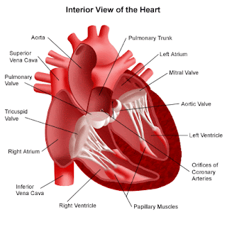
Human heart is cone shaped,hollow muscular structure that lies in the thoracic cavity above the diaphragm and in between two lungs.Heart is completely enclosed with a thin walled double layered pericardium.Between the double layered membrane,pericardial fluid is found which protects the heart from friction and keep the heart moist.It also protect the heart from mechanical injuries.
Human heart is four chambered organ with two auricles and two ventricles.The transverse groove distinctly separates the heart into anterior auricular part and posterior ventricular part which is the largest part of the heart.The auricles are thin walled separated from each other by an inter auricular septum whereas the ventricles are thick walled separated from each other by an oblique inter ventricular septum.In the upper region of right auricle,near the precaval opening there is presence of Sinu Auriculo node or SA node through which heart beat starts and is also called as natural pacemaker of heart.The right and left auricles open into their respective ventricles through Auriculo ventricular aperture.The right Auriculo ventricular aperture is guarded by tricuspid valve while the left auriculo ventricular aperture is guarded by bicuspid or mitral valve.These valve are attached with papillary muscles of columnae cornae of the ventricles through chordae tendinae,which prevent the valves from being pushed into auricles at the time of ventricular contraction.The precavals and post caval open into the right auricle through separate openings.Two pulmonary vein open in the left auricle by a common opening.A pulmonary trunk arises from the right ventricle which carry deoxygenated blood to the lungs for oxygenation.An aortic trunk arises from left ventricle which carries oxygenated blood to the different parts of the body.The opening of pulmonary and aortic trunk is guarded by three semilunar valve.
WORKING OF HEART:
The heart collects the blood through both the auricles and distributes it through the ventricles,The action of heart includes the contraction and relaxation of auricles and ventricles.The rhythmic contraction and relaxation of heart is know as heart beat.The contraction of heart is known as systole and the relaxation of heart is known as diastole.The contraction and relaxation of different parts of heart take place in a definite order.Auricles and ventricles contract alternately.
The right auricles receive the deoxygenated blood from the body through two main vessel :the precavals(collect the impure blood from the head and upper parts of the body)And post cavals(from lower part). Impure blood is pumped into right ventricle due to contraction of right auricle.When the right ventricle contract,deoxygenated blood is pushed through pulmonary artery to the for purification.The left auricle receives pure blood from the right and left pulmonary veins.The blood is passed through the corresponding left ventricles.The left ventricle contracts as a result of which aorta pumps oxygenated blood and sent to different parts of the body.

ORIGIN OF HUMAN HEART BEAT:

The rhythmic contraction and relaxation of heart is known as heart beat.The heart beat begins in the tiny island of tissue in the upper region of right auricle at the point of precaval opening called Sinu auricular node or SA node.This region is also called the natural pacemaker as Heartbeat initiated from it.A wave of contraction(Systole)originates from the region and spreads to both auricles which causes its contraction.At the posterior part of right auricle i.e at the junction of right auricle and venticle,a tissue called auricula ventricular node or AV node is present which picks the wave of contraction propagated by SA node.Av node divide into left and right branches that extends the wall of ventricles on corresponding side and reach upto the apex called Bundle of His.This fibre form the network which spread on the entire wall of ventricle called Purkinge fibre.The excitation wave from AV node spreads along Bundle of His and Purkinge fibres ,and thus excites the muscle of ventricles to contract simultaneously.

HUMAN HEART SOUNDS:

Heart sounds are the sounds that are produced due to certain closure of the valves of heart.It is accomplished by two heart sounds.The first heart sound indicates the beginning of Ventricular systole.It is also called LUBB.It is produced due to the closer of Auriculo ventricular valve.The second sound or DUBB indicates the end of ventricular systole and beginning of its diastole.It is produced due to closure of semilunar valve.
First sound----Second sound----pause

TYPES OF HEART:

1)Myogenic heart:
The heart in which wave of contraction starts from muscle fibres of hearts are said to be Myogenic Heart.
Eg:Human heart
2)Neurogenic Heart:
the hearts in which wave of contraction starts from nerve cells or group of such cells are said to be neurogenic heart.
Eg:Cray fish heart
ARTIFICIAL PACE MAKER:
When the sinu auricular node becomes defective or damaged the heart beat becomes irregular and abnormally slow downs.This problem can be rectified by surgical grafting of an instrument known as Artificial pacemaker.It was first implanted by Chardack in 1960.The implanted pacemaker raises the heart beat rate to normal.The pacemaker is made of pulse generator and an electrode.The pulse generator is a sealed box containing lithium halide cells to generate power for more than ten years and an electric circuit to regulate the heart beat rate.The electrode is a fine metallic string which is connected to the pulse generator.Its special tip remains in contact with the interior right ventricle.
The pacemaker is placed under the collar bone below the skin by a simple operation.A pacemaker can be replaced or removed easily.

Plants Anatomy along with secondary Growth

VASCULAR BUNDLE:
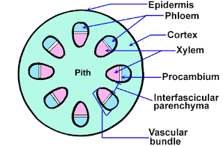
The bundle which consists of tissue like xylem and phloem is known as vascular bundle.

TYPES OF VASCULAR BUNDLES:
1)RADIALS:
The vascular bundle in which xylem and phloem are found in different radius is known as radial vascular bundle.It is the characteristics feature of root.
Eg:monocot and dicot root

2)CONJOINT:
Conjoint vascular bundle is a type of vascular bundle in which xylem and phloem are found in same radius or bundle.
Eg dicot or monocot stem
a)COLLATERAL:
This is the condition in which xylem lies towards innerside and phloem towards outer.
i)CLOSED COLLATERAL:
In this condition,cambium is absent.
Eg:monocot stem
ii)OPEN COLLATERAL:
In this condition,cambium is present.
Eg:dicot stem

b)BICOLLATERAL:
This is the condition where the phloem and cambium lie on both sides of the xylem.
Eg stem of cucurbita family
3)CONCENTRIC:
It is a type of vascular bundle where the xylem and phloem are found in concentric rings.It is of following type:
a)AMPHICRIBAL/HADROCENTRIC:
This is the condition where the xylem lies in the centre and phloem surrounds it.
Eg:fern
b)AMPHIVASAL/LEPTOCENTRIC:
This is the condition where the phloem lies in the centre and xylem surround it.
Eg:Yucca,dracaena
Technique:amphicirbal----Bahira----phloem
Amphivasal-----vitra----phloem
PATTERN SHOWN BY XYLEM:
TECHNIQUE:Take protoxylem as reference and define.

1)EXARCH:
Pattern shown by xylem in which protoxylem lies towards periphery and metaxylem towards centre is known as exarch.
Eg:Monocot and dicot root

2)ENDARCH:
Pattern shown by xylem in which protoxylem lies towards the centre and metaxylem towards the periphery is called endarch.
Eg:monocot and dicot stem

3)MESARCH:
The condition in which protoxylem lies middle of metaxylem is knowm as mesarch.
Eg: Rachis and rhizome of certain fern

TERMS:
1)INTRAFASICULAR CAMBIUM:Cambium of vascular bundle
2)INTERFASICULAR CAMBIUM:Cambium between the vascular cambium
PLANTS ANATOMY:
1)ANATOMY OF DICOT ROOT:Plants Anatomy along with secondary Growth
The T.S. of dicot root shows the following internal structure listed below:
a)EPIBLEMA:
Epiblema is the outermost single layer compactly arranged and bears numerous root hairs but without cuticle.It is parenchymatous in nature.

b)CORTEX:
Cortex lies just below the epiblema which consists of several layer of parenchymatous tissue with intercelluar spaces.These tissues help in storage of food materials.

c)ENDODERMIS:
Endodermis is the single layered parenchymatous tissues with barrel shape and compactly arranged cells.the lateral walls becomes thick and waxy which prevents the flow of water and minerals to form Casparian Stripes.Some cells having thin wall lie just against the protoplasm through which water and mineral are passed into stellar rPlants Anatomy along with secondary Growthegion is called as passage cells.

d)PERICYCLE:
Pericycle is single layer lying just below the endodermis.It helps to develop lateral roots,secondary meristem and cork cambium.
e)CONJUCTIVE TISSUE:
These are thin walled tissue that separates xylem and phloem in vascular bundle.
f)VASCULAR CAMBIUM:
Vascular cambium consists of xylem and phloem which lie on different radii i.e is of radial type.The protoxylem lies towards the periphery whereas metaxylem lies towards the center;thus xylem is of exarch.Xylem is composed of tracheids and vessels whereas phloem consits of sieve tube,companion cells and phloem parenchyma.The main function of these vascular element is conduction of mineral and water(xylem);and food material(phloem).Vascular bundles ranges from 2-6.
g)PITH:
Pith is centrally located groups of parenchymatous tissue.In dicot roots,it may be absent but if present is found in reduced form due to secondary growth.

2)ANATOMY OF MONOCOT ROOT:
The T.S. of monocot root shows the following internal structure listPlants Anatomy along with secondary Growthed below:
a)EPIBLEMA:
Epiblema is the outermost single layer compactly arranged and bears numerous root hairs but without cuticle.It is parenchymatous in nature.

b)CORTEX:
Cortex lies just below the epiblema which consists of several layer of parenchymatous tissue with intercelluar spaces.These tissues help in storage of food materials.A few layer of cortex just below the epidermis dies off when they get older and cutinize to form exodermis.

c)ENDODERMIS:
Endodermis is the single layered parenchymatous tissues with barrel shape and compactly arranged cells.the lateral walls becomes thick and waxy which prevents the flow of water and minerals to form Casparian Stripes.Some cells having thin wall lie just against the protoplasm through which water and mineral are passed into stellar region is called as passage cells.
d)PERICYCLE:
Pericycle is single layered structure in the form of ring lying just below the endodermis.It is parenchymatous in nature and helps to develop lateral roots.

e)CONJUCTIVE TISSUE:Plants Anatomy along with secondary Growth
These are thin walled tissue that separates xylem and phloem in vascular bundle.

f)VASCULAR CAMBIUM:
Vascular cambium consists of xylem and phloem which lie on different radii i.e is of radial type.The protoxylem lies towards the periphery whereas metaxylem lies towards the center;thus xylem is of exarch.Xylem is composed of tracheids and vessels whereas phloem consits of sieve tube,companion cells and phloem parenchyma.The main function of these vascular element is conduction of mineral and water(xylem);and food material(phloem).Vascular bundles ranges from 6-20.

g)PITH:
Pith is centrally located groups of parenchymatous tissue and is well developed.

3)ANATOMY OF DICOT STEM:
The T.S. of dicot stem shows the following internal structure:
a)EPIDERMIS:
Epidermis is the outermost layer which is made up of parenchymatous Plants Anatomy along with secondary Growthcells compactly arranged and consisting numerous hairs.It is covered by cuticle which helps to check excess transpiration and external injuries.

b)HYPODERMIS:
Below epidermis is few layers of collenchymatous cell called hypodermis.It helps in mechanical function.

c)CORTEX:
A immediate layer below hypodermis is made up of few layer of parenchymatous cells consisting intercellular space called cortex.It helps in storage of food.

d)ENDODERMIS:
Endodermis lie just below the cortex and is made up of single layer of parenchymatous cell which are barrel shaped and compactly arranged.It consists of starch grain and is also known as starch sheath.
e)PERICYCLE:
Pericycle lies just below the endodermis and consists of 2-3 layers of parencymatous or schlerchymatous tissue.

f)VASCULAR BUNDLES:Plants Anatomy along with secondary Growth
The vascular bundle which is in the form of ring consists of xylem and phloem.It is of conjoint,colateral and open type i.e xylem and phloem are separted by cambium.The protoxylem lies toward the centre whereas metaxylem towards the periphery.Thus the xylem is endarch.Xylem consists of tracheids,vessels,wood fibre and xylem parenchyma whereas phloem consists of sieve tube,companion cells ,phloem parenchyma and bast fibre.These vascular tissue helps in the conduction of water,minerals and synthesized food.

g)PITH:
Pith is centrally located group of parencymatous cells that helps to store food.It is well developed.

h)MEDULLARY RAYS:
Group of elongated parenchymatous cell that extend from pith to cortex region in between two vascular bundles is called medullary rays.It helps to exchange food material.

4)ANATOMY OF MONOCOT STEM:
The T.S of monocot stem shows the following internal tissue listed below:
a)EPIDERMIS:
Epidermis is the outermost layer which is made up of parenchymatous cells compactly arranged and is covered by cuticle which helps to check excess transpiration and external injuries.

b)HYPODERMIS:
Below epidermis is few layers of sclerenchymatous cell called hypodermis.It helps in mechanical function.

c)GROUND TISSUE:
Ground tissue is multilayered,parencymatous that extends from hypodermis to the centre of stem.It is not differentiated into cortex and stellar region(endodermis,pericycle and vascular bundle).Plants Anatomy along with secondary Growth

d)VASCULAR BUNDLES:
Vascular bundles are foind in scattered form.The smaller bundles are distributed towards the pheriphery while the larger distributed towards centre.Each vascular bundle consist of xylem and phloem covered by bundle sheath.The vascular bundle is conjoint,colateral and closed type(Remember mC3)i.e absence of cambium.The xylem is endarch.The xylem consists of large vessel known as metaxylem and smaller protoxylem called lysigenous cavity formed by the disintegration of some protoxylem elements.The phloem consists of sieve tube,companion cells and bast fibre.these vascular tissue helps in the conduction of water,mineral and food material.

e)PITH:
Pith is completely absent.

NOTE:Phloem parenchyma is absent in monocot stem.


SECONDARY GROWTH:
The increase in thickness or diameter of plant axis due to formation of secondary tissue by the activity of cork or vascular cambium in the extra stellar or stellar region is known as secondary growth.Secondary growth doesn't take place in Monocot stem due to absence of cambium except in Yucca,Dracaena which are exception.
PROCESS OF SECONDARY GROWTH: 1)FORMATION OF COMPLETE RING OF VASCULAR CAMBIUM:
some of the medullary rays cells mostly in the line of intrafasicular cambium becomes meristematic and forms a strip of interfasicular cambium.The intrafasicular cambium joins with interfasicular cambium forming a complete ring of vascular cambium known as cambium ring.

2)ACTIVITY OF VASCULAR CAMBIUM:
The vascular cambium consists of single row of ray initial and fusiform initials.Plants Anatomy along with secondary Growth
Ray initial forms parenchymatous medullary rays whereas fusiform initials form vascular elements xylem towards inner side and phloem toward outer side due to more activeness of vascular cambium in inner side than outer side.Fusiform initial divide periclinically giving rise to tissue mother cell which differentiate into secondary xylem cell inside the cambium and into secondary phloem outside the cambium.
In primary stem,primary xylem and primary phloem are nearer but due to formation of secondary xylem and phloem,they are shifted apart.Primary phloem are in the form of patches but later on they are crushed whereas primary xylem remain till the end in small amount at pith.
3)ANNUAL RING:
The activity of vascular cambium is not uniform due to climatic condition.The vascular cambium is more active during spring and is called spring wood whereas it is less active in autumn and is called autumn wood.Secondary xylem is more and larger in nPlants Anatomy along with secondary Growthumber in spring wood rather than in autumn wood.One spring wood and autumn wood forms annual ring.By counting the number of annual rings ,age of that particular part can be predicted and this process is known as dendrochronolgy.

4)SAP WOOD AND HEART WOOD:
The light colour wood formed on the outer side of tree in the late year which takes part in conduction is known as sap wood or alburminum.It is made of living as well as dead cell and is poor quality of wood.
The dark coloured wood formed on inner side of tree in the early year is known as heart wood or duranum.It is made of dead cell which is thickened.thus it does notPlants Anatomy along with secondary Growth take part in conduction.

5)ORIGIN AND ACTIVITY OF CORK CAMBIUM:
A strip of secondary meristem called cork cambium or phellogen commonly originates in the outer layer of the cortex.The activity of cork cambium gives up new cells on both sides forming the secondary cortex(phelloderm) towards the inner side and cork(phellem) on outer side.the secondary cortex generally contains chloroplast and carries photosynthesis while the cork found outside the cork cambium is roughly rectangular and consists of suberin which is impervious to water.That's why it is used in bottle cork.
BARK:
Bark is dead tissue lying outside the active cork cambium which include epidermis,lenticels and the cork.It function is to provide protection.
LENTICELS:
The aerating pores formed in bark through which exchange of gases take place are called lenticels.
SIGNIFICANCE OF SECONDARY GROWTH:
1)It provides support to increasing weight of aerial growth.
2)It produces corky bark and protects the interior part.
3)It adds new conducting tissue for replacement of old non functioning tissue.